

Click on the images below to work through three patient cases and learn more about the signs and symptoms of FOP that can help with establishing a prompt, correct diagnosis and avoiding inadvertent harm.
Click on the profiles to discover more:

About
5-year-old Brandon presents with a swelling on his back. The swelling appeared after he fell from a swing and was painful and hot to the touch. His parents began to worry when it did not go down after a few days and Brandon became feverish.1
1. Pignolo RJ et al. J Bone Miner Res 2016;31:650–656

Patient history
- Brandon developed jaundice two days after birth, which was resolved by the time he was two weeks old
- Brandon’s parents reported that his big toes were shorter than his others and turned inwards towards the rest of his toes, but were told this would likely correct itself as he grew
- Brandon’s parents visited the pediatrician when he was 18 months old as he had not yet learned to crawl but would instead shuffle on his bottom. The pediatrician told them that all children developed at different rates, and Brandon began to walk just after his 2nd birthday
- Brandon was treated for bronchiolitis at two years old

Which factors of Brandon’s history would lead you to suspect a diagnosis of FOP?
Based on the swelling on Brandon’s back and his history, FOP is suspected. How can a diagnosis of FOP be confirmed?

Diagnosis

About
Jade first visited a doctor at seven years old with a large, tender swelling in her right shoulder that appeared aſter a school gym class.
An MRI identified patterns of calcification within the lesion and a diagnosis of traumatic myositis ossificans was made. Jade was prescribed non-steroidal anti-inflammatory drugs and advised to ice the bump and rest until the swelling went down.1
Click on the text below to learn more about these early signs of FOP
Myositis ossificans ![]()
Myositis ossificansMyositis ossificans (MO) is a rare disorder characterized by the growth of heterotopic bone in skeletal muscle and surrounding soft tissue. It can be hereditary, non-traumatic (associated with burns, hemophilia, and neurological conditions) and traumatic (associated with direct or repetitive trauma). Traumatic MO is the most common and usually occurs due to sport-related impact.1
MO usually presents as a warm, tender swelling that progresses to a palpable mass, and symptoms include joint and muscle stiffness, pain and decreased range of motion.1 Swelling, pain, and joint stiffness are also symptoms of FOP flare-ups, making MO a common misdiagnosis in patients with FOP.2–4

Second diagnosis
Six months later, Jade’s swelling had not subsided, and she still had limited movement in her shoulder. Her parents took her to a different doctor when a similar, painful swelling appeared in her upper back.
The doctor disagreed with her previous diagnosis and, aſter examining her hands and feet, made a new diagnosis of FOP.2
Click on the text below to learn more about these early signs of FOP
Signs of FOP ![]()
 Image from Acharya S et al, 20184
Signs of FOPClassic FOP is defined by two clinical features:
Malformation of the great toes and progressive heterotopic ossification. Thus, a definitive diagnosis of FOP can be based on the presence of rapidly appearing soft tissue lesions in addition to great toe malformations.1
Other developmental anomalies may also be present, including:
- Short-bone: Shortened thumbs, shortening of the second phalanx of the fiſth finger, short broad femoral neck2
- Cervical spine: Large posterior elements, tall, narrow vertebral bodies, fusion of the facet joints between C2 and C73
1. Pignolo RJ et al. Pediatr Endocrinol Rev 2013;10:437–448; 2. Thickman et al. AJR 1982;139:935–941; 3. Schaffer AA et al. Spine 2005;30:1379–1385; 4. Acharya S et al. J Nepal Health Res Counc 2018;16:245–247.
Image from Acharya S et al, 20184
Approximately what percentage of patients with FOP are initially misdiagnosed?


About
At 14 months old, Ricky’s parents visited their pediatrician as they had noticed a rapidly growing, immobile nodule on Ricky’s scalp. The nodule appeared spontaneously and did not appear to cause him any pain.1 The pediatrician suspected that the nodule could be malignant and referred Ricky to oncology.2
1. Piram M et al. J Am Acad Dermatol 2011;64:97–101; 2. Kaplan FS et al. Proc Intl Clin Council FOP 2022;1:2–127.
Could the scalp nodule have been a symptom of FOP?

Biopsy
Ricky’s oncologist performed a diagnostic excisional biopsy of the nodule, which ruled out malignancy.1
However, the skin at the biopsy site thickened and hardened, and a new nodule occurred.1,2 Ricky’s pediatrician was concerned that the biopsy had worsened his condition and consulted with a colleague.

Consultation
The colleague reviewed Ricky’s case and suggested a diagnosis of FOP. They highlighted the presence of great toe malformations that are indicative of FOP and should always be examined in an infant with scalp nodules.2
Click on the text below to learn more
Great toe malformation ![]()
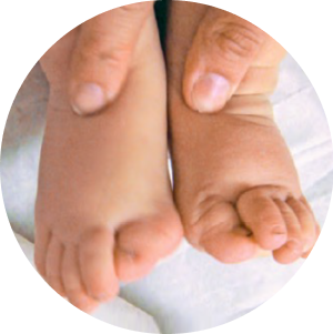 Image from Asadi S et al, 20205
Great toe malformationThe presence of scalp nodules during infancy should prompt immediate examination of the great toes.1
Almost all patients with FOP have congenital bilateral great toe malformations,2,3 which include hallux valgus, shortened great toes and sharpening of the first metatarsal bone.4
Identification of great toe malformations in patients with scalp nodules can accelerate the proper diagnosis of FOP.1
Image from Asadi S et al, 20205
The consulting doctor explained that biopsies should not be performed in patients with FOP, as they are likely to cause additional heterotopic ossification.2
Ricky’s parents were advised that certain other activities/procedures can also cause flare-ups of FOP and should be avoided.
Click on the text below to find out more
What to avoid in patients with FOP ![]()
Activities and procedures to avoidCertain activities can cause flare-ups and should be avoided or prevented where possible:1,2

Over-exertion or tiredness

Surgery

Certain dental procedures

Biopsy

Intramuscular injections (such as vaccinations)

Muscular stretching and passive range of motion exercises

Injuries, such as bumps, bruises and falls

Future considerations
Ricky’s diagnosis was confirmed via a genetic test, which revealed a c.617G>A (p.R206H) mutation in ACVR1/ALK2.3
Ricky’s parents are trying for another baby and are concerned about the risk of future children having the same mutation. Neither of Ricky’s parents show any signs or symptoms of FOP.
1. Piram M et al. J Am Acad Dermatol 2011;64:97–101; 2. Kaplan FS et al. Proc Intl Clin Council FOP 2022;1:2–127;
3. Shore EM et al. Nat Gen 2006;38:525–527.
